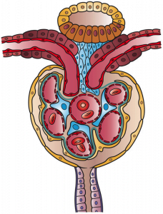
It was long believed that the kidney is a “static” organ once it develops, but researchers in Israel and America recently discovered that, contrary to popular belief, the human kidney is capable of regenerating itself.
The study, conducted by researchers at Sheba Medical Center, Tel Aviv University, and Stanford University is set to change the way we think about our kidneys by identifying the precise cellular signaling responsible for kidney regeneration and exposing the multifaceted nature of kidney growth.
Kidney regeneration: not just confined to petri dishes
Such a discovery is revolutionary in the medical field as kidney regeneration could potentially serve as an alternative to kidney transplants. In the United States, the Center for Disease Control (CDC) estimates that nearly 10 percent of adults – more than 20 million people – suffer from chronic kidney disease, which is a condition in which the kidneys are damaged and cannot filter blood as effectively as healthy kidneys.
Dr. Benjamin Dekel, the principal researcher at Tel Aviv University, began researching the subject of kidney regeneration three years ago while on sabbatical at Stanford University. Though the laboratory experiments and stem cell research were conducted at Stanford, the results were analyzed by researchers at TAU and Stanford.
Related articles:
- NaNose: The Breathalyzer Test That Sniffs Out Lung Cancer Before It Spreads
- Meet The 6 Israeli Startups On ‘Forbes’ Top Ten Health Tech Changing The World
Previously, scientists knew kidney cells could reproduce outside the body, but the biological process taking place inside the body at the single cell level was never explored. Uncovering that process became the focus of Dr. Dekel’s efforts: “We wanted to change the way people thought about kidneys — about internal organs altogether,” said Dr. Dekel. “Very little is known even now about the way our internal organs function at the single cell level. This study flips the paradigm that kidney cells are static — in fact, kidney cells are continuously growing, all the time.”
Discovering the fountain of kidney life
The researchers conducted the study using a “rainbow mouse” model developed at Stanford’s Weissman lab. A “rainbow mouse” is a genetically engineered mouse designed to express one of four fluorescent markers called “reporters” in each cell, which allowed the researchers to follow cell growth inside of a living mammal. Kidney growth, they were surprised to find, was sectional and multi-directional. What this means is that the building blocks of the kidney, known as nephrons, have multiple parts that each grow at a different pace and in a different direction, like the branches of a tree.
Using the mouse, they were also able to pinpoint a specific molecule responsible for kidney cellular growth called the “WNT signal.” Once activated in precursor cells, the WNT signal results in robust cellular growth and generation of long branches of cells.According to Dr. Dekel, “No one had ever used a rainbow mouse model to monitor development of kidney cells. It was exciting to use these genetic tricks to discover that cellular growth was occurring all the time in the kidney — that, in fact, the kidney was constantly remodeling itself in a very specific mode.”
Now armed with this discovery of kidney regeneration in mice, the next step is to achieve kidney regeneration in humans. This could lead to alleviating the shortage of kidney organs for transplantation. “If we were able to further activate the WNT pathway, then in cases of disease or trauma we could activate the phenomena for growth and really boost kidney regeneration to help patients,” said Dr. Derkel.
The research, published in Cell Reports, was conducted by Dr. Dekel of TAU’s Sackler School of Medicine and Sheba Medical Center and Dr. Irving L. Weissman of Stanford University’s School of Medicine, working with teams of researchers from both universities.
Photos: Medical Imaging – Male Organs – Kidneys, BigStock/ Holly Fischer
